
Risks of NOT Getting a Vasectomy
If a couple wants to have a child, the risks associated with reversible forms of contraception to delay pregnancy until they are ready, and the risks associated with the ultimate…
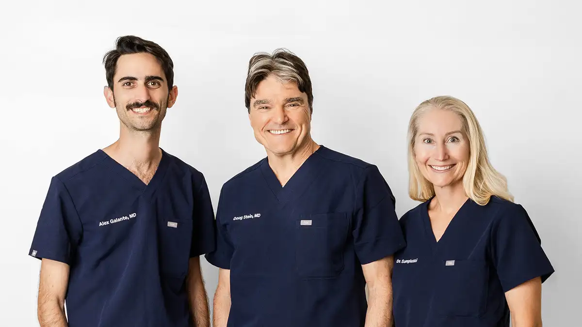
Our entire practice is dedicated solely to vasectomies and reversals. This super-subspecialized approach allows us to offer a level of experience, consistency, and technical precision rarely found in general urology practices.
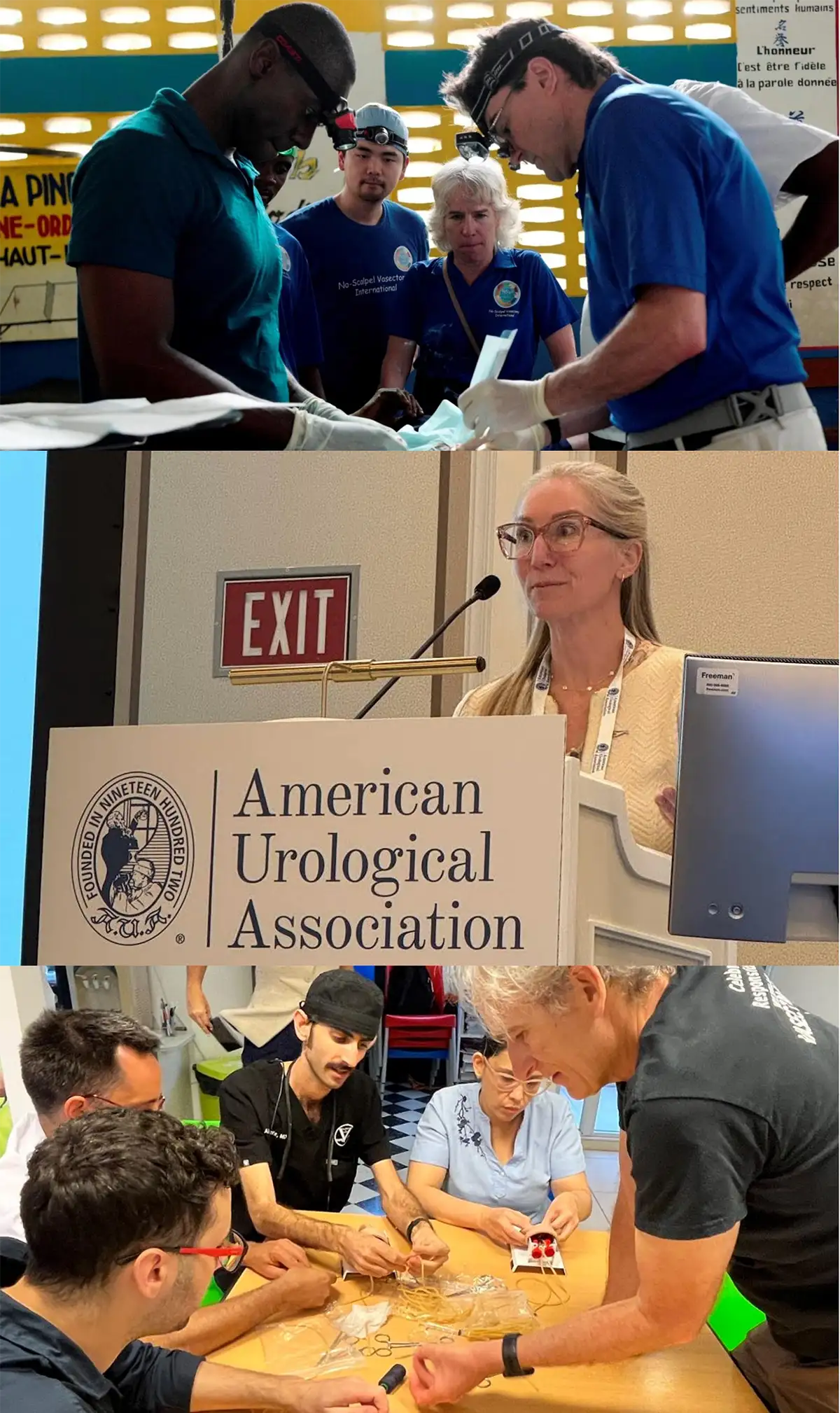
We have been focused exclusively on vasectomies and reversals since 2001. Our physicians have performed tens of thousands of procedures and are among the most experienced vasectomy specialists in the world. We also train doctors from across the U.S. and internationally in minimally invasive, highly effective vasectomy techniques, and we participate in global volunteer programs to expand access to safe family-planning care.
Most patients can be seen within 1–2 weeks, not months. When you call us, a human will answer the phone. We combine the consultation and the procedure into a single visit, minimizing time away from work and family.
Before your appointment, you review our website and watch our counseling video at your own pace. This makes the in-office experience smoother, faster, and more personalized to your specific questions/concerns.
We accept most insurance plans, and our self-pay pricing is highly competitive. We offer discounts through training programs and county health departments.
Every patient receives their doctor’s personal cell phone number for postoperative questions or concerns. This level of availability is uncommon and reflects our commitment to excellent care.
We are board-certified urologists. Men from all over the country—and the world—have chosen our practice. Many have shared their experiences on Google, Reddit, and other public platforms. Do your own research. We’re known for our light-hearted billboards, but we take our medical care extremely seriously. We emphasize evidence-based technique, safety, comfort, efficiency, and clear communication.
Often covered by health insurance & Title X
Not Covered by Insurance